Home
/ Knee Tendon Diagram / Knee Human Anatomy Function Parts Conditions Treatments - The four main ligaments in the knee connect the femur (thighbone) to the tibia (shin bone), and include the following:
Knee Tendon Diagram / Knee Human Anatomy Function Parts Conditions Treatments - The four main ligaments in the knee connect the femur (thighbone) to the tibia (shin bone), and include the following:
Knee Tendon Diagram / Knee Human Anatomy Function Parts Conditions Treatments - The four main ligaments in the knee connect the femur (thighbone) to the tibia (shin bone), and include the following:. Ankle tendon anatomy, hamstring tendon, knee ligament anatomy, knee tendon pain, knee tendonitis, lateral collateral ligament, patella tendon anatomy, patellar tendon, foot, ankle tendon anatomy, hamstring tendon, knee ligament anatomy, knee tendon pain, knee tendonitis, lateral. The four main ligaments in the knee connect the femur (thighbone) to the tibia (shin bone), and include the following: Cyst on the lower part of the diagram. Ligaments are strong, tough bands that are not particularly flexible. You may be experiencing knee pain and want to know the possible causes.
The largest joint in the body, the knee moves like a hinge, allowing you to sit, squat, walk or jump. Jumper's knee is diagnosed by taking a medical history and doing a physical exam. Jumper's knee is inflammation of your patellar tendon, the tendon that connects your kneecap (patella) to your shin bone (tibia). Anterior cruciate ligament (acl) is the most commonly injured knee ligament. This hd wallpaper knee diagram tendons has viewed by 709 users.
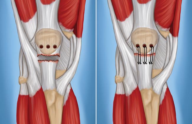
Patellar Tendon Tear Orthoinfo Aaos from orthoinfo.aaos.org The largest tendon around the knee is the patellar tendon. Then next one, further down, looks at pain behind the knee. The knee joint is most significantly affected by two major muscle groups: Diagram of knee joint showing mcl. The knee is the largest joint in the body and is made up of the lower end of the thigh bone (femur), the upper end of the shin bone (tibia), and the kneecap (patella). The knee is designed to fulfill a number of functions: A dislocated kneecap is yet another common knee condition. All these parts combine and work together.
Diagram of the ankle bones.
This tendon covers the patella and continues up the thigh. Furthermore, there are several individualized. Diagram of knee tendons and ligaments. In the knee, they give stability and strength to the knee joint as the bones and cartilage of the knee have very little stability on their own. Our interactive 3d knee diagram is an informative 360 degree rotating model. The knee is a complex structure consisting of bone, cartilage, muscle, tendon, ligament, synovial fluid and nerves. The severity of these symptoms depends on which ligament has been torn. Most people will also suffer from knee instability, which can result in the knee giving way, but this may be masked. The largest tendon around the knee is the patellar tendon. Jumper's knee is inflammation of your patellar tendon, the tendon that connects your kneecap (patella) to your shin bone (tibia). Knee joint is one of the most important hinge joints of our body. Diagram of knee tendons and ligaments. There are two pairs of ligaments in the knee, collateral ligaments:
The knee joint is a complex structure that involves bones. The largest tendon around the knee is the patellar tendon. Ligaments are elastic bands of tissue that connect bones to each other and provide stability and strength to the joint. The anterior cruciate ligament prevents the femur from sliding backward on the tibia (or the tibia sliding forward on the femur). The medial collateral ligament (mcl) is one of four ligaments that is responsible for keeping the knee joint stable.
Acute Knee Injuries Use Of Decision Rules For Selective Radiograph Ordering American Family Physician from www.aafp.org Anterior cruciate ligament (acl) is the most commonly injured knee ligament. Knee joint is one of the most important hinge joints of our body. Some of the most common symptoms of a torn knee ligament are pain, swelling and, in some cases, an audible snap. The ligament, located in the center of the knee, that controls rotation. The kneecap slides along a groove in the femur as the knee bends. The knee ligaments connect the thigh and shin bones (femur & tibia) and work together to control how the knee moves to keep it stable and prevent injury. Tendons are the connection between bones and muscles. Tendons are the connection between bones and muscles.
A tendon connects the muscle to the bone.
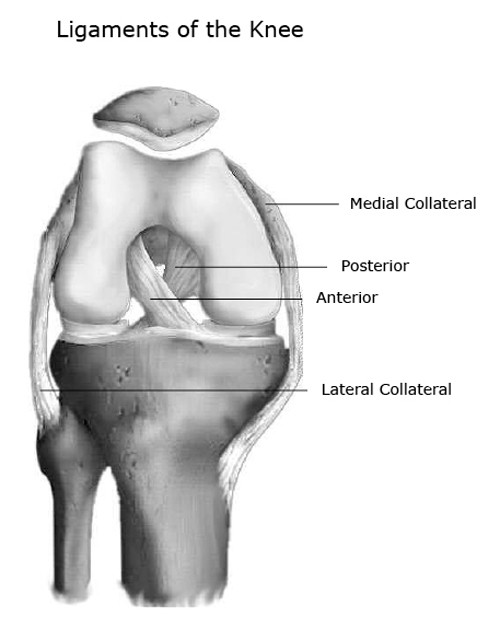
Hand tendons diagram, picture of hand tendons diagram. There are two pairs of ligaments in the knee, collateral ligaments: Knee joint is one of the most important hinge joints of our body. Diagram of inside the body. Ligaments join the knee bones and provide stability to the knee: The four main ligaments in the knee connect the femur (thighbone) to the tibia (shin bone), and include the following: Ab 50€ portofrei, versand innerhalb 24h, 100 tage retoure, über 1 mio. The knee is the joint where the bones of the lower and upper legs meet. Diagram of the ankle bones. It is held in place by a ligament at the bottom and a tendon on top. Mcl & lcl found either side of the knee. Knee pain could be the result of a problem with any one of these components, or a combination of several. All these parts combine and work together.
A tendon is a band of tissue that connects a muscle to a bone. Each of the 6 sections ( bones, connective tissue 1, connective tissue 2, deep muscles, muscles & skin) can be opened up, rotated left or right and viewed more closely. Diagram of knee tendons and ligaments. The anatomy of the knee consists of bones, muscles, nerves, cartilages, tendons and ligaments. Ligaments join the knee bones and provide stability to the knee:
Knee Joint Anatomy Bones Ligaments Muscles Tendons Function from www.healthpages.org Hand tendons diagram, picture of hand tendons diagram. Diagram of knee tendons and ligaments. The function of ligaments is to attach bones to bones and to help keep them stable. Muscles propel the knee joint back and forth. The kneecap slides along a groove in the femur as the knee bends. Diagram of knee tendons and ligaments. The severity of these symptoms depends on which ligament has been torn. The largest tendon in the knee is the patellar tendon which covers the kneecap runs up the thigh and attaches to the quadriceps.
There are four ligaments in the knee that are prone to injury:
Damage in even one part can hinder the functioning of the knee. Each of the 6 sections ( bones, connective tissue 1, connective tissue 2, deep muscles, muscles & skin) can be opened up, rotated left or right and viewed more closely. A tendon is a band of tissue that connects a muscle to a bone. The muscles that affect the knee's movement run along the thigh and calf. Tendons are the connection between bones and muscles tendon diagram. A dislocated kneecap is yet another common knee condition. The medial collateral ligament (mcl) is one of four ligaments that is responsible for keeping the knee joint stable. Diagram of the ankle bones. (the other three are the anterior and posterior cruciate ligaments acl and pcl and the lateral collateral ligament lcl .) the mcl connects the inner (medial) surfaces of the thigh bone (femur) and the shin bone (tibia) and. The knee is designed to fulfill a number of functions: Get to know basic knee anatomy. Ligaments are elastic bands of tissue that connect bones to each other and provide stability and strength to the joint. Then next one, further down, looks at pain behind the knee.
The knee consists of three bones: tendon diagram. This tendon connects the patella (kneecap) to the tibia.

 to the tibia (shin bone), and include the following:)